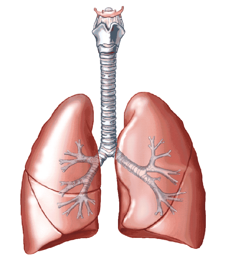
LOS ANGELES: Scientists, including those of Indian origin, have successfully grown three-dimensional (3D) lungs in the lab, using stem cells, which can be used to study diseases that are difficult to understand with conventional methods.
By coating tiny gel beads with lung-derived stem cells and then allowing them to self-assemble into the shapes of the air sacs found in human lungs, researchers created 3D “organoids”.
The laboratory-grown tissue can be used to study diseases including idiopathic pulmonary fibrosis, which has been difficult to study using conventional methods, researchers said.
“While we haven’t built a fully functional lung, we’ve been able to take lung cells and place them in the correct geometrical spacing and pattern to mimic a human lung,” said Brigitte Gomperts, an associate professor at University of California, Los Angeles (UCLA) in the US.
Idiopathic pulmonary fibrosis is a chronic lung disease characterised by scarring of the lungs.
The scarring makes the lungs thick and stiff, which over time results in progressively worsening shortness of breath and lack of oxygen to the brain and vital organs.
After diagnosis, most people with the disease live about three to five years. Though researchers do not know what causes idiopathic pulmonary fibrosis in all cases, for a small percentage of people it runs in their families.
Additionally, cigarette smoking and exposure to certain types of dust can increase the risk of developing the disease.
To study the effect of genetic mutations or drugs on lung cells, researchers have previously relied on 2D cultures of the cells.
However when they take cells from people with idiopathic pulmonary fibrosis and grow them on these flat cultures, the cells appear healthy.
The inability to model idiopathic pulmonary fibrosis in the laboratory makes it difficult to study the biology of the disease and design possible treatments.
Researchers, including Manash Paul, Saravanan Karumbayaram and Preethi Vijayaraj from UCLA, started with stem cells created using cells from adult lungs.
They used those cells to coat sticky hydrogel beads, and then they partitioned these beads into small wells, each only seven millimetres across.
Inside each well, the lung cells grew around the beads, which linked them and formed an evenly distributed three-dimensional pattern.
To show that these tiny organoids mimicked the structure of actual lungs, the researchers compared the lab-grown tissues with real sections of human lung.
When researchers added certain molecular factors to the 3D cultures, the lungs developed scars similar to those seen in people who have idiopathic pulmonary fibrosis, something that could not be accomplished using 2D cultures of these cells.
Using the new lung organoids, researchers will be able to study the biological underpinnings of lung diseases including idiopathic pulmonary fibrosis, and also test possible treatments for the diseases. (AGENCIES)


