Dr M K Mam
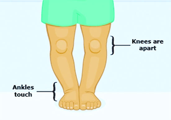
Bow legs – genu varum is a very common deformity found in infants and toddlers. There is bowing of the legs as the legs are curved outwards at the knees while child’s feet are together. As such knees remain apart when child stands with feet and toes pointing forwards, there is a space between the knees and lower legs. The deformity is usually bilateral, however at times the deformity can be found in one leg only.
Causes: The bowing can be a) physiological b) pathological
a) Physiological bow legs: Bow legs is normal in babies due to their folded position in the mother’s womb. It is considered as a normal stage of development and gets corrected slowly as the child grows. Bowing usually becomes prominent when child starts standing and walking. It is usually bilateral, mild in nature and is symmetrical. The deformity corrects naturally in most of the children. It begins to correct slowly at around 12 to 18 months of age and continues to improve. Bowing is corrected fully and legs are normal by 3-4 years of age. When the bow legs continues and the legs do not straighten by 3 to 4 years of age or is getting worse or is severe , we need to rule out pathological cause for the bowing.
b) Pathological bow legs: The bowing is because of pathology- disease resulting in disturbance of the growth at the knee. Any disturbance in the growth plate i.e. the epiphysis especially of the inner part of the upper end of the shin bone i.e. tibia or the lower end of thigh bone i.e. femur produces bowing of legs, as the growth is disturbed in inner part whereas it is taking place normally in outer part of growth plate. Pathological bowing is usually asymmetrical and does not improve with the passage of time. It usually requires bracing and / or corrective surgery. Various conditions which can cause pathological bowing include-
i) Metabolic disorders like Vitamin D deficiency in children causes rickets. Bones become soft and weak in rickets which results not only in bow legs but also deformities in other bones. Nutritional rickets has been a very common cause of bowing in developing countries where mal and under nutrition is a common problem. Rickets can also be caused by a genetic abnormality that does not allow Vitamin D to be absorbed correctly.
ii) Blount’s disease results in a common type of pathological bow legs. It is a growth disorder where there is abnormality of growth of the upper end of shin bone in inner and back part of growth plate. The deformity is progressive and gets worse over the time. It is bilateral in majority of patients. These children are usually overweight and have a family history of bow legs. An early diagnosis and treatment of the condition is essential as that helps in improving the prognosis and reducing the complications.
iii) Injury to the inner part of growth plate of upper end of shin bone or lower end of thigh bone can disturb growth in the inner part when normal growth is taking place in the outer part of the growth plate and the result is bowing. The bow leg caused by injury is usually unilateral.
iv) Any pathology like infection or tumour of inner part of the growth plate of upper end of shin bone or lower end of thigh bone damages the inner part of the growth plate and this results in bowing of legs as normal growth is going on outer part of the growth plate. Again, pathological bow leg due to infection or tumour is usually unilateral.
v) Bone dysplasia also produces bowing as there is disturbance in growth plate.
vi) Achondroplasia, a bone growth disorder results in dwarfism and there can also be bowlegs.
Clinical picture: Bow legs is the complaint that the parents present the child with. It becomes very evident when child stands and walks. When a child with bow legs stands with feet together toes pointing forward, ankles may touch but the knees remain apart. It usually does not bother the child as it does not cause any discomfort or pain. The child with physiological bow legs can usually crawl, walk, run, play like a child of his age. However, the parents are worried about the deformity- the appearance of the child’s legs, or awkward walking pattern of child. When it is on one side only, child can have a functional limb length discrepancy. Persistent bowing in adolescents can result in pain or discomfort in hips, knees and ankles because of abnormal stresses that the bowed legs have on these joints. The severity of bow legs- deformity can be assessed by measuring the amount of distance between the knees or the angle between leg and thigh when the child stands or lies down with the feet together.
Diagnosis: Diagnosis of bow legs is made by taking a detailed history and physical examination. Relevant blood tests are done to rule out any vitamin D or calcium deficiency. X-Rays of legs are done in standing position to rule out any pathology in the bones especially any problem in growth of the bones. X-Rays also help in assessing the location and extent of the deformity.
Treatment: Physiological bowing in toddlers and babies usually does not need any active treatment unless it is severe. However, these children need to be regularly followed – assessed at 6 monthly intervals for the progress – resolution of the deformity. The parents as usual are very much concerned, as such we should educate, explain and reassure them. Exercises are important to strengthen the muscles and bones, however they do not change the shape of bones. Modification of shoes like slight shoes raise on the outer side of the shoes are being prescribed in children with mild degree of deformity. Braces i.e. knee -ankle -foot orthosis are also suggested, they may be helpful in correcting the deformity slowly. However, there are reports that corrective shoes or braces are not of much help, rather they may hinder the normal straightening of legs. Braces are helpful in early cases of Blount’s disease and bowing because of rickets. In children with rickets, vitamin D and calcium has to be given in addition to braces. When the deformity is of severe degree or when there is an underlying problem like Blount’s disease , it needs surgical correction. One option of surgery is ‘guided growth’ wherein we temporarily stop the growth on the healthy outer part of the upper end of shinbone by putting a staple or small plate, thereby give a chance to abnormal inner side to catch up and over the time the leg shall straighten in opposite direction with the child’s natural growth. The staple or plate is removed once the deformity is corrected. Other option is to do a corrective osteotomy wherein the bone is surgically divided at the upper end of shin bone or lower end of thigh bone and repositioned to correct the deformity. It is usually done in severe cases or in the cases without growth remaining to allow guided growth to be successful. Surgery is usually deferred until near or at the end of the growth i.e. skeletal maturity (unless it is severe),because there is a risk of recurrence of the deformity as child is still growing. Surgery as it is, has to be meticulously planned considering all factors like age of the child – bone age, the expected further growth etc.
(The author is Formerly, Vice Principal , Prof. and Head Orthopaedics, Christian Medical College, Ludhiana, Punjab. )
Trending Now
E-Paper


