Dr Arvind Kohli
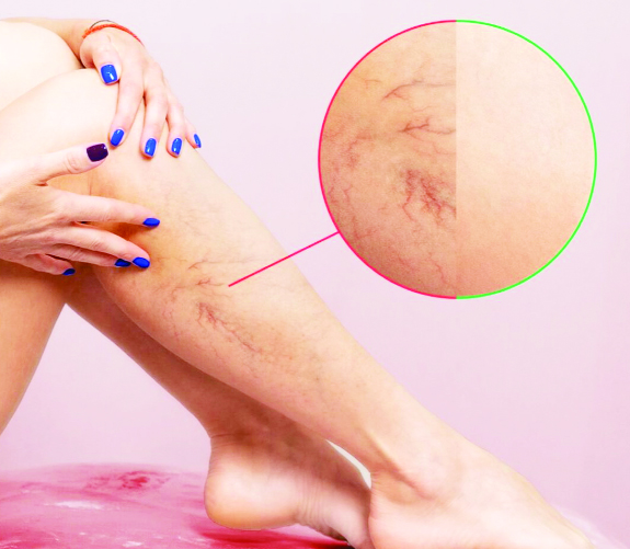
Varicose veins are veins that have become swollen enough to be seen on the legs. This condition affects the superficial veins, which lie closest to the skin. The affected veins appear as blue bulging, twisted lumps under the skin. Varicose veins are commonly found among people who stand on their feet a lot. It’s a condition that can run in the family and affects as many as 25 million Indians (16% of men and 29% of women)
Varicose veins appear when excess blood pools in the superficial veins of the leg. . Varicose veins are twisted, dilated veins most commonly located on the lower extremities. Risk factors include chronic cough, constipation, family history of venous disease, female sex, obesity, older age, pregnancy, and prolonged standing. The exact pathophysiology is debated, but it involves a genetic predisposition, incompetent valves, weakened vascular walls, and increased intravenous pressure.
Similar to varicose veins seen with superficial veins, chronic venous insufficiency (CVI) is a condition that occurs when blood pools in the superficial and deep leg veins. CVI can occur with or without the presence of varicose veins. This condition develops when the blood pressure in the veins is abnormally high. CVI can occur after veins have been damaged by injuries or blood clots. People with CVI often have a combination of symptoms.
Symptoms and signs of CVI include Restless leg syndrome (pain, cramping in legs) hyperpigmentation, stasis dermatitis, , chronic edema and venous ulcers.
* Edema begins in the perimalleolar region and ascends up the leg.
* Sense of discomfort in legs is often referred to as weight or pain after standing for a long time and it is relieved by leg elevation.
* Cutaneous changes include skin hyperpigmentation with hemosiderin deposition and eczematous dermatitis.
* This fibrotic process produces lipodermatosclerosis and there are risks of cellulites, leg ulcers and delayed wound healing.
* In addition, chronic venous insufficiency contributes to the development of lymphedema
* What is Atrophie Blanche?
This condition manifests as small porcelain-white scars resulting from skin micro-infarction in an area affected by venous hypertension, caused by the obstruction of dermal arterioles and subsequent infarction (death of the tissue due to obstruction of the blood supply) of the skin supplied by these blood vessels.
Management of Varicose Veins New advances have changed the practice of venous interventions Varicose Vein treatment has evolved into an interventional therapy, in part due to the new technologies available.and most recent are being explained
Wearing compression stockings: The most conservative approach is to wear properly-fitting compression stockings. Compression stockings with pressure gradient steadily squeeze the legs, helping veins and leg muscles circulate blood more efficiently., preventing venous ulcers and accelerating wound healing process while alleviating cramping and calf pain
Surgical Treatment Traditional surgical treatment consisted of ligation and stripping of the greater saphenous vein with avulsion of tributary veins(phlebectomies).
Thermal Ablation
Thermal ablation for superficial truncal reflux has revolutionized vein therapies in the 21st century. though selected patients may still be better treated with ligation (with or without phlebectomy). Thermal ablation can be performed with endovenous laser ablation (EVLA) or radiofrequency ablation (RFA).
EVLA Endovenous laser ablation has revolutionized the management of varicose veins. The laser wavelength can target water or hemoglobin, leading to thermal injury to the target vein endothelium and its resultant occlusion. The EVLA fiber is a low-profile device that can be advanced over a wire and then activated and withdrawn slowly. Target veins can include an incompetent great saphenous vein (GSV), small saphenous vein (SSV), accessory, or perforator. Using ultrasound (US) guidance, a sheath is placed into the target vein, typically as downstream as feasible. The fiber tip, with US guidance, is placed at least 2 cm downstream from the deep vein junction. .. After the careful administration of tumescent anesthesia, ablation can proceed.Patient can be discharged shortly after the procedure
Radiofrequency ablation (RFA) is another thermal-based technology to occlude saphenous veins.. An electrode element is inserted into the target vein. The tissue acts as a resistor, and the molecules surrounding the element become excited and heat up. Thermal injury occurs within the vein lumen leading to thrombosis and eventual occlusion . Just as with EVLA, tumescent anesthesia is administered around the target vein. Tumescent anesthesia contains saline, lidocaine, epinephrine, and bicarbonate. .
NTNT Non-Thermal Techniques Though thermal techniques have shown good efficacy for sealing incompetent veins, they have some shortcomings. They require the application of tumescent anesthesia, which involves additional needle insertions for its application. This leads to a prolonged procedure time and is somewhat painful part of the procedure for the patient Several non-thermal, non-tumescent (NTNT) techniques to eliminate axial reflux and varicose veins are now available for use. These currently include Ultrasound guided Foam sclerotherapy, &Mechanochemical ablation (MOCA),
MOCA induces endothelial inflammation, thrombosis, and occlusion by combining mechanical injury and chemical irritation. The mechanical action of the spinning fiber is also thought to induce venoconstriction allowing for greater sclerosant contact with the endothelium. The device has a battery within its motorized handle. No generator is requiredMechanical rotation is initiated for at least 3 s together with slight withdrawal of the fiber without the administration of sclerosant, to induce venospasm and reduce entry of the sclerosant into the deep venous system..
Glue Another available technology to seal the saphenous veins is cyanoacrylate adhesive (“superglue”) Various cyanoacrylate formulations have been used in medicine for decades. Through a sheath, a delivery catheter is advanced into the saphenous vein. Through the catheter, the cyanoacrylate is carefully injected using a gun-handle mechanism. Within the GSV, typically a 5-cm distance is observed from the SFJ . to prevent glue migration into the deep system. After every administration using ultrasonic visualization, the catheter is withdrawn, and prolonged manual pressure is applied. After the entire desired length of vein has been treated, the fiber and sheath are removed. Compression or wraps are not necessary.
Why Treat them early Varicose Veins are just one aspect of a larger issue and can often lead to various Complications (especially DVT and venous ulcers) And Prompt diagnosis and treatment of Varicose Veins can help prevent many of these complications to happen.
Current evidence shows that management of venous diseases is now shifting towards minimally invasive interventions with very promising results Keeping in view the increasing incidence of pathological venous diseases these procedures have revolutionized the management of Varicose Veins have become standard of practice offered for patients with chronic venous disease.
(The author is Vascular Surgeon SVMM Hospital Jammu)
Trending Now
E-Paper


